


Criteria for diagnosis
Osteoarthritis of the limbs
The radiographic diagnosis of osteoarthritis is based on 4 signs that may be isolated or associated with one another:
- Localised joint space narrowing, usually asymmetrical. This primarily reflects a reduction in thickness of the cartilage and its deterioration. It is necessary to assess where it is localised and how significant it is.
- Marginal osteophytes
- Epiphyseal osteocondensation
- Epiphyseal or "hyper-pressure" geodes, rounded or oval size. They are most often located in the area of epiphyseal osteocondensation of the subchondral bone.
The first two signs are sufficient to consolidate a diagnosis often sensed clinically.
In the event of joint space narrowing without osteophytes the diagnosis may be more difficult and require additional imaging examinations.
Spinal osteoarthritis
Discarthrosis results in overall or localised narrowing of the intervertebral disc space, subchondral condensation and osteophytes on the vertebral endplates. Zygapophysial osteoarthritis is revealed by condensation of the facet joints, joint space narrowing on three-quarter views, osteophytes, remodelling and horizontalization of joint surfaces, sometimes responsible with disc degeneration of spondylolisthesis.
In most cases, standard radiography remains the appropriate tool for the diagnosis of osteoarthritis.
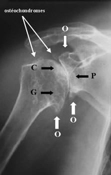
O : Osteophyte - P : Joint space narrowing - G : Geode - C : Compression
Major form of shoulder osteoarthritis (omarthrosis) with stage IV omobrachial joint space narrowing, exuberant osteophyte formation in the lower section of the humeral head and neck of the scapula, osteosclerosis and osteochondromas
CH Lyon Sud - Pr. Vignon, Dr. Conrozier et Dr. Mathieu
Other interests in standard radiography
Search for the aetiological factors of osteoarthritis
The X-rays are used to find the aetiological factors of osteoarthritis such as dysplasia for the hip, chondrocalcinosis, traumatic after-effects, etc. and signs of complications such as secondary synovial osteochondromatosis.
Monitoring the structural evolution
Standard radiography also enables monitoring the structural evolution and thus constitutes one of the elements of the prognosis of the osteoarthritic disease. Structural monitoring is done by X-ray every 18 to 24 months. Measuring joint space is used to evaluate narrowing.
Repeated inflammatory flare-ups of osteoarthritis in the lower limbs may be related to a rapid chondrolysis. Close radiological monitoring may therefore be necessary.
In standard radiography several scores enable classifying patients according to the severity of their illness. The Kellgren and Lawrence grading scale is the most commonly used. This is an index that takes into account the osteophytes and joint space narrowing. It comprises 4 grades: doubtful, moderate, definite and severe osteoarthritis.
Grade 0 corresponds to a normal X-ray and grade 4 to complete loss of cartilage. This type of scoring can be used to assess the progression of osteoarthritis and can be applied to many joints.
Major principles of standard radiography
There are some key principles of standard osteoarticular radiography:
One must always X-ray the two symmetrical joints in order to compare the pathological side to the asymptomatic side presumed healthy.
Indeed, when the X-ray reveals an abnormal image, if it is bilateral, it may be an anomaly with no clinical significance.
In addition, taking an X-ray of two symmetrical joints sometimes enables discovering an bilateral impairment that is asymptomatic on one side. This scenario is relatively common for example in hip osteoarthritis.
In the lower limbs (especially the knee), the X-rays should be made in a position of constraint, preferably supporting body weight. Indeed, in a standing position, the joint space narrows a little more because of the stress to the skeleton imposed by the weight of the body. Potential narrowing may fade completely when the body is relieved of body weight and mislead the diagnosis.
One must always do X-rays based on several views to better assess the joint space but by limiting the views actually needed to minimise the amount of exposure delivered to the patient.
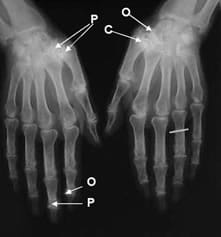
O : Osteophyte - P : Joint space narrowing - C : Condensation (sclerosis)
Bilateral digital osteoarthritis affecting the distal interphalangeal joints and bilateral rhizarthrosis
CHU Pitié-Salpétrière - Dr. Gibert
Limitations of standard radiography
Typically, radiological signs are present in the early clinical manifestations of osteoarthritis, but X-rays may be normal or subnormal in the early stages of the disease due to the slow development of radiographic signs. The clinician can then make use of modern imaging techniques, which can provide structural and lesional information and enable appropriate early treatment.
Early diagnosis of osteoarthritis is interesting because of the possibilities of preventive surgery and the hopes that future structure-modulating drugs may bring.
In this category
-
 Diagnosis
Clinical examination
-
 Diagnosis
Other imaging examinations