


Pain is the most common reason for consultation.
Generally located in the groin, it may radiate to the front of the thigh or anteromedial thigh. External pain with regard to the greater trochanter, and posterior pain radiating to the back of the thigh are less evocative.
Painful irradiations can continue down to the knee.
This is a mechanical pain, increased during exertion, relieved by rest, and not waking the patient up at night (except for changes in position in very advanced stages).
It may be accompanied during inflammatory episodes by a feeling of morning stiffness, a stiffness lasting less than 15 minutes.
The pain may be accompanied by an impact on daily activities and walking, with reduced walking distance and discomfort during the rotation of the hip (difficulty putting on socks, pulling on a pair of tights).
Algofunctional indexes (such as those of Lequesne and WOMAC) are used to assess the degree of functional impairment.
An examination of the hip must be carried out first of all standing, then walking, and finally lying down.
In a standing position one looks for lameness of the hip, a vicious attitude in hip flexion or in external rotation.
In a lying down position on finds pain and/or a painful limitation in mobilisation of the hip. In the early stages movements are somewhat limited and the limitation is on internal rotation and abduction. It is worth noting the presence of quadriceps amyotrophy.
Pitfalls to avoid
- Metabolic arthropathy
- A non-osteoarticular pathology: crural neuralgia, crural hernia, femoral arteritis and phlebitis, etc.
- Bone disease: hip trauma, Paget's disease of the pelvis, osteonecrosis of the femoral head, etc.
- Abarticular pathology: gluteal tendinitis of the right anterior, etc.
In this category
-
 Clinical examination
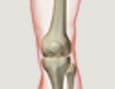
Knee osteoarthritis
-
 Clinical examination
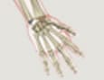
Osteoarthritis of the hands
-
 Clinical examination
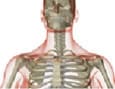
Cervical osteoarthritis
-
 Clinical examination
Lumbar osteoarthritis
-
 Clinical examination
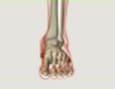
Osteoarthritis of the feet